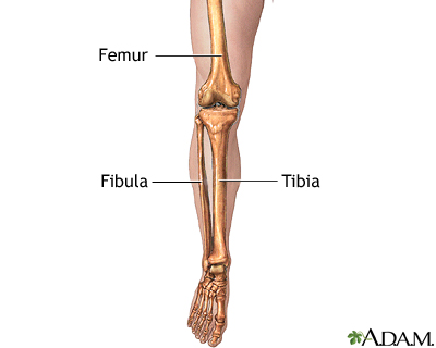
Lower Leg Bones Diagram - Leg Definition Bones Muscles Facts Britannica : The proximal portion of the tibia is tibial plateau which acts as a cusp for the knee, the distal portion tapers into the medial malleoli and the concave surface which articulates with the talus at the ankle joint.
Lower Leg Bones Diagram - Leg Definition Bones Muscles Facts Britannica : The proximal portion of the tibia is tibial plateau which acts as a cusp for the knee, the distal portion tapers into the medial malleoli and the concave surface which articulates with the talus at the ankle joint.. This diagram depicts lower leg bones 1024×1350.human anatomy diagrams show internal organs, cells, systems, conditions, symptoms and sickness information and/or tips for healthy living. The femur, or thighbone, is the longest and largest bone in the human body. Its lower end helps create the knee joint. The foot bones shown in this diagram are the talus, navicular, cuneiform, cuboid, metatarsals and. The lower leg is made up of two very strong, long bone—the tibia and the fibula.
Ebraheim's educational animated video describes the muscle and nerve anatomy of the lower leg.there are fourteen muscles within the lower leg. Legs are used for standing, and all forms of. Start studying lower leg bones. Learn vocabulary, terms, and more with flashcards, games, and other study tools. While their parts are similar in general, their structure has been adapted to differing functions.
Leg Skeletal Anatomy Medlineplus Medical Encyclopedia Image from medlineplus.gov It is located toward the middle of the lower leg. The bones of the leg are the femur, tibia, fibula and patella.the foot bones shown in this diagram are the talus, navicular, cuneiform, cuboid, metatarsals and calcaneus. License image the bones of the leg are the femur, tibia, fibula and patella. The femur, or thighbone, is the longest and largest bone in the human body. Legs are used for standing, and all forms of. Our goal is that these leg anatomy worksheets pictures gallery can be a direction for you, bring you more references and also make you have a great day. The fibula, or calf bone, is smaller and is located on the outside of the lower leg. Diagram of blood and nerve supply to bone.
Distal to the ankle is the foot.
#diagram and names of leg bones #diagram of foot and leg bones #diagram of leg bones #diagram of lower leg bones #diagram of the bones in your leg related posts of diagram of leg bones inside of arm muscle and bone Our goal is that these leg anatomy worksheets pictures gallery can be a direction for you, bring you more references and also make you have a great day. The lower leg is comprised of two bones, the tibia and the smaller fibula. The lower leg is made up of two very strong, long bone—the tibia and the fibula. At the same time, the bones and joints of the leg and foot must be strong enough to support the body's weight while remaining. The lower leg is also home to nerve fibers. The talus the weight of your body is transferred from the tiba to the talus. The fibula is the smaller shin bone that runs down the outer side of the lower leg. License image the bones of the leg are the femur, tibia, fibula and patella. While their parts are similar in general, their structure has been adapted to differing functions. These are the femur, patella, tibia, fibula, tarsal bones, metatarsal bones, and phalanges (see figure 6.51). It is located toward the middle of the lower leg. Distal to the ankle is the foot.
#diagram and names of leg bones #diagram of foot and leg bones #diagram of leg bones #diagram of lower leg bones #diagram of the bones in your leg related posts of diagram of leg bones inside of arm muscle and bone This area is commonly referred to as the calf. Develop an understanding of the causes of equine lameness and methods of treatment. The hip bone (os coxae, innominate bone, pelvic bone or coxal bone) is a large irregular bone, constricted in the center and expanded above and below leg bone diagram. The femur, or thighbone, is the longest and largest bone in the human body.
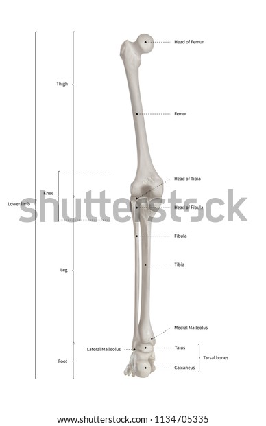
Infographic Diagram Human Skeleton Lower Limb Stock Illustration 1134705335 from image.shutterstock.com This allows weight to be distributed either anteriorly or posteriorly throughout the foot. The talus the weight of your body is transferred from the tiba to the talus. Diagram of blood and nerve supply to bone. The femur, or thighbone, is the longest and largest bone in the human body. They connect the lower leg to the rest of the body and gives stability, flexibility and strength. Bone diagram forehead (frontal bone) nose bones (nasals) cheek bone (zygoma) upper jaw (maxilla) lower jaw (mandible) breast bone (sternum) upper arm bone (humerus) lower arm bone (ulna) thigh bone (femur) collar bone (clavicle) toe bones (phalanges) ankle bones (tarsals) kneecap (patella) shin bone (tibia) calf bone (fibula) foot bones This area is commonly referred to as the calf. Anterior muscles of the lower leg, lateral fibularis group and posterior muscles of the lower le.
Riesige auswahl an cds, vinyl und mp3s.
The ligament joining the two bones of the lower leg (tibia and fibula), called the syndesmotic ligament, is injured. Click now to learn more about the bones, muscles, and soft tissues tibia: The muscles of the lower leg can divided into 3 main groups: A high ankle sprain causes pain and swelling similar to a. At the bottom it also expands outwards, forming a bony prominence known as the lateral malleolus. While their parts are similar in general, their structure has been adapted to differing functions. Ebraheim's educational animated video describes the muscle and nerve anatomy of the lower leg.there are fourteen muscles within the lower leg. The musculoskeletal system including bones muscles, cartilage, tendons, ligaments and joints; Any disorder or defect in the knee bone can restrict the activities of the leg which can directly affect our locomotion. Also called the shin bone, the tibia is the longer of the two bones in the. The bones of the leg and foot form part of the appendicular skeleton that supports the many muscles of the lower limbs. They connect the lower leg to the rest of the body and gives stability, flexibility and strength. This area is commonly referred to as the calf.
The muscles of the lower leg, called simply the leg by anatomists, largely move the foot and toes. The medial, larger bone of the lower leg. The lower leg extends from the knee to the ankle. Riesige auswahl an cds, vinyl und mp3s. Lower leg pain is common, but it can be tricky sorting out its many potential causes.
Lippincott Williams Wilkins Atlas Of Anatomy Musculature Chart Lower Limb Anatomical Chart Company 9781605471051 from d1w7fb2mkkr3kw.cloudfront.net Many muscles that move the trunk and legs, such as our abdominal muscles, attach to the hip bones. It extends slightly lower than the medial malleolus forming the outer side of the ankle joint where it articulates with (meets) the talus. The thigh bone, or femur, is the large upper leg bone that connects the lower leg bones (knee joint) to the pelvic bone (hip joint). Our goal is that these leg anatomy worksheets pictures gallery can be a direction for you, bring you more references and also make you have a great day. At the bottom it also expands outwards, forming a bony prominence known as the lateral malleolus. Its lower end helps create the knee joint. The lower leg is comprised of two bones, the tibia and the smaller fibula. The talus the weight of your body is transferred from the tiba to the talus.
The fibula, or calf bone, is smaller and is located on the outside of the lower leg.
Lower leg pain is common, but it can be tricky sorting out its many potential causes. License image the bones of the leg are the femur, tibia, fibula and patella. The hip bone (os coxae, innominate bone, pelvic bone or coxal bone) is a large irregular bone, constricted in the center and expanded above and below leg bone diagram. The femur, or thighbone, is the longest and largest bone in the human body. The musculoskeletal system including bones muscles, cartilage, tendons, ligaments and joints; The medial, larger bone of the lower leg. Diagram of blood and nerve supply to bone. The bones of the leg are the femur, tibia, fibula and patella.the foot bones shown in this diagram are the talus, navicular, cuneiform, cuboid, metatarsals and calcaneus. The fibula, or calf bone, is smaller and is located on the outside of the lower leg. The femur, or thighbone, is the longest and largest bone in the human body. Ebraheim's educational animated video describes the muscle and nerve anatomy of the lower leg.there are fourteen muscles within the lower leg. Damage to the nervous system. Anterior muscles of the lower leg, lateral fibularis group and posterior muscles of the lower le.
The fibula is the smaller shin bone that runs down the outer side of the lower leg leg bones diagram. The largest and most medial leg bone, forming both the knee and ankle joints.


0 Komentar